Thursday 09 / April / 2026
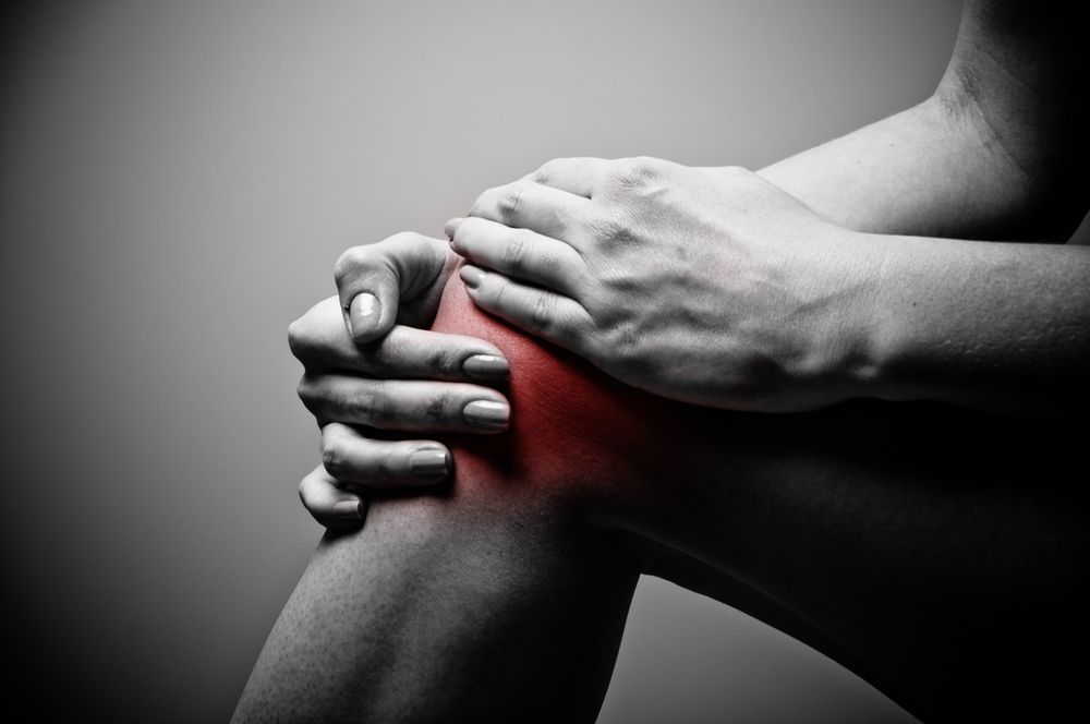
Osteoarthritis (OA) in the knee is defined as a degeneration of joint cartilage and the underlying supporting structures in normal joints. A firm, rubbery material called cartilage covers the end of each bone. Cartilage provides a smooth, gliding surface for joint motion and acts as a cushion between the bones. In OA, the cartilage breaks down, causing pain, swelling, and problems moving the joint. In the body, an inflammatory process occurs and cytokines (proteins) and enzymes develop that further damage the cartilage. In the final stages of OA, the cartilage wears away and bone rubs against bone leading to joint damage and more pain. Although OA occurs in people of all ages, osteoarthritis is most common in people older than 65. Common risk factors include increasing age, obesity, and previous joint injury, overuse of the joint, weak thigh muscles (quadriceps), and genetic predisposition. Occupational habits are also known to be a risk factor, with the risk of knee OA significantly elevated in those whose job involves more than 30 minutes per day squatting, kneeling, or climbing stairs. Other pre disposing conditions are gout (increased uric acid level), rheumatoid arthritis, and Paget’s disease of the bone.
Prevalence of Osteoarthritis
It is one of the top five most disabling conditions that affects more than one-third of persons above 65 years of age. Global estimates reveal more than 100 million people are affected by OA. The financial expenditures for the care of persons with OA are estimated at a total annual national cost estimate of $15.5-$28.6 billion per year. The COPCORD studies conducted in India, Bangladesh, and Pakistan looked specifically into differences between rural and urban populations. In India the crude prevalence of clinically-diagnosed knee OA was higher in the urban (5.5%) than the rural community (3.3%).
Symptoms in OA knee:
• Pain
• Stiffness — more severe on waking up in the morning, and improves within 30 minutes when the individual starts moving about
• Swelling
• Deformity
Diagnosis
• Physical examination
• Painful range of motion
• Muscle strength in the affected region
• Presence of any swelling or tenderness of the joint
• Gait (the way you walk) if OA is in the hip or knee
• Joint line tenderness
• Radiological examination
• X-rays are very helpful in diagnosing osteoarthritis because the affected joint will have a characteristic appearance, such as:
• Bones appearing closer to each other: As cartilage wears away, the joint space can narrow.
• Cysts: As the body responds to cartilage destruction and attempts to stabilise the joint, cysts or fluid-filled cavities can form in the bone.
Increased bone density or uneven joints: When bones are no longer cushioned by cartilage, they can rub against one another, creating friction. The body responds by laying down more bone in response, increasing bone density. Increased bone creates uneven joint surfaces and osteophytes (bone spurs) around the joint margins. MRI (magnetic resonance imaging) MRI can demonstrate reactive bone edema or soft tissue swelling as well as small cartilage or bone fragments in the joint. CT (computed tomography) examinations are excellent for demonstrating the degree of osteophyte (bone spur) formation and its relationship to the adjacent soft tissues. CT examinations are also useful in providing guidance for therapeutic and diagnostic procedures. Ultrasound is extremely sensitive for identifying synovial cysts that can form in association with osteoarthritis. Ultrasound can also be used to image articular cartilage in patients who cannot tolerate an MRI examination because of claustrophobia or pacemakers. Bone scans are very sensitive in detecting reactive bone turnover association with osteoarthritis. Bone scans can also image the entire skeleton in one examination and thus can provide the clinician with helpful information in patients in whom there may be multiple areas where OA is present.
Laboratory findings: Laboratory tests are helpful in the diagnosis of OA because they are usually normal. Routine tests such as complete blood counts, urinalysis, sedimentation rate (ESR), biochemistries, and specialised tests such as rheumatoid factor and antinuclear antibody (ANA) are useful simply to rule out other diseases that cause joint pain. However, it should be remembered that as we age, a low level positive test for rheumatoid arthritis (rheumatoid factor) or ANA, and elevations of sedimentation rate (ESR) can sometimes develop without the presence of the illness. Although these findings can sometimes be confusing to a patient, they need not be. The clinical picture makes the diagnosis; lab tests are used only to confirm the clinical picture. Laboratory tests should never be used alone to diagnose arthritis. Synovial fluid is the liquid that is normally found within the joints. It helps nourish and lubricate the joints. It is usually present in only very small amounts. However when arthritis is present, it changes in character and amount. Withdrawing the fluid can reduce swelling and pain. It can also help to confirm the diagnosis. When the synovial fluid is removed, it should be sent for culture, as well as be tested for cell count. In osteoarthritis, the white cell count (“pus cells”) is usually low and the fluid is clear (like water); higher counts should suggest inflammatory arthritis or infection. The fluid may also be examined for the presence of uric acid crystals (seen in gout) or calcium pyrophosphate crystals. The measurement of other biological markers is still experimental.
Treatment Modalities
Weight reduction is one of the first and unproblematic measures that can be taken to reduce knee OA. Studies of OA have constantly shown that overweight people have higher rates of knee OA than non-overweight control subjects. This is due to the fact that the force across the knees is 3-6 times the body weight; therefore, people who have more mass cause extreme forces on their knees, leading to the early onset or steady progression of knee OA condition. The most widely used remedy for knee OA is rehabilitation and physical therapy (PT). PT has proved to be useful in helping patients with pain and mobility. Fitness walking, aerobic exercise, and strength training have all been reported to result in functional improvement in patients with OA of the knee.
Strength training, being the most common treatment approach for the management of patients with functional limitations, is prescribed to address the need to increase muscular strength and joint stability for improving in WOMAC (Western Ontario and Mc-Master Universities OA Index) pain scores and overall health benefits. The common equipment used is based on fundamentally different movement progression and resistance patterns such as isotonic (unchanged tension but change in length), isometric (no change in length or angle), and isokinetic (constant resistance with variation in speeds). Though isometric activities show effective results in reducing pain levels, they are avoided when working with the elderly due to increases seen in heart rate and blood pressure, which could be contraindicative to other co-morbidities.
Hydrotherapy (balneotherapy) involves the use of water in any form or at any temperature (steam, liquid, ice) for the purpose of healing. Hydrotherapy/balneotherapy and aquatic therapy displayed positive results, when conducted for testing subject’s strength and flexibility. The sessions typically are run from six to 48 weeks for a duration of 60 minutes, and are conducted in a shallow pool with water temperatures ranging from 29°C to 34°C. Patients are typically recommended to exercise between 50% and 70% target heart rate for a minimum of 30 minutes, thrice a week, for overall weight management, health benefits, and a reduction in pain which was noted after a six-month programme.
Yoga’s gentle movements can aid to build body strength, flexibility, and balance, and reduce arthritis pain and stiffness. The slow, controlled physical movement of joints is helpful for the arthritis patients. It improves the blood circulation in joints, removing unwanted toxins and other waste products. A pilot study conducted by the University of Pennsylvania, School of Medicine, examined one type of yoga, Iyengar Yoga, suitable for people with OA of the knee. After an eight-week course of weekly 90-minute beginner classes, there was a statistically significant reduction in pain, physical function, and mood, indicating the positive effects of yoga therapy for OA rehabilitation.
Heat/cold therapy: Heat, cold, pressure, light, and even electricity have been used for thousands of years to accelerate healing and decrease pain. Transcutaneous electrical nerve stimulation (TENS) is one of the most widely used physical modalities for the management of OA knee. Many studies have already been completed and demonstrated no significant differences in immediate pain relief between the groups.
Pharmacological agents: Acetaminophen, aspirin, and non-steroidal anti-inflammatory drugs (NSAIDs) are commonly used as pain relief medicines to treat OA. Excessive use of NSAIDs can lead to gastric complications, ulcers, increased risk for hospitalisation and other adverse side effects. Glucosamine sulphate and chondroitin sulphate are two nutritional supplements that have been reported to “cure” arthritis. As glucosamine and chondroitin are produced within the body and are used in the manufacture or repair of cartilage, it is suggested that the synthetic versions work the same way.
Intra articular injections of hyaluronic acid and steroids: Hyaluronic acid is the lubricating substance within the joint which may be lost during osteoarthritis. It has been approved by the US Food and Drug Administration as a device for the treatment of OA in humans since 1997.
Total joint arthroplasty: Generally with a knee replacement, a metal cap is fitted on to the bottom of the thigh bone (femur) and a plate is fitted on top of the shin bone (tibia). The joint between the two is a plastic hinge joint. As against the common myth that total knee patients are not satisfied with the surgical outcomes, total hip and total knee arthroplasties were found to be quite effective in terms of improvement in health-related quality-of-life dimensions. Age was not found to be an obstacle to effective surgery, and men seemed to benefit more from the intervention than did women. When improvement was found to be modest, the role of comorbidities was highlighted.
Unicondylar joint replacement: It is most common for arthritis problems to affect the medial (inside) aspect of the knee; therefore, partial knee replacements can be done and only the inner portion of the knee joint is replaced. When replacing the medial knee, more range of motion is expected along with faster recovery.
Arthroscopic knee debridement (key hole surgery): The small inconsequential benefit seen from interventions that include arthroscopy for the degenerative knee is limited in time and absent at one to two years after surgery. Knee arthroscopy is associated with harms in patients with OA. Taken together, these findings do not support the practice of arthroscopic surgery for the middle aged or older patients with knee pain with or without signs of osteoarthritis. Currently, arthroscopic partial meniscectomy and/or loose body removal is routinely performed in patients with symptomatic OA of the knee who also have primary signs and symptoms of a torn meniscus and/or a loose body.
In conclusion, the primary goals of the management of patients with OA are pain control and to bring improvement in function and health-related quality of life, with avoidance of toxic pharmacological effects. Total knee replacement is indicated in patients with severe osteoarthritis suffering extensive pain and deformity, where conservative measures have failed. Obesity is not a contra indication for total joint replacement against the myth among most patients and there is no difference in patient reported outcome measures in obese patients, although wound complications were significantly higher. Patient satisfaction is generally improved in obese patients following total knee replacement and therefore, it should be utilised in severe disease after discussing the relative benefits and risks of surgery. —lifestyle@timesofoman.com
Dr Raghavan Sivaram is specialist orthopaedic surgeon at Apollo Hospital, Muscat